Almost every area of modern biology, from molecular to planetary scales, is being revolutionized by quantitative experiments. At the same time, developments in statistical mechanics and dynamical systems and our exponentially expanding computational abilities have prepared the physics community to address questions posed by more complex systems[1]. What once was far too complicated and difficult to measure or predict now is, or may soon be, within the realm of questions simple enough to be usefully addressed by physics, mathematics, and computation.
At the same time scientific and engineering disciplines of every kind are focusing ever more closely on the atomic and molecular properties of the world around us[2]. This shift in scientific study generally goes under the name of nanotechnology, though much of it is science not technology at this point.
Study of the small has driven the development of new experimental techniques and instruments, one of the most successful being the family of instruments known as Scanning Probe Microscopes (SPM). Though these types instruments are now in some cases decades old, new and interesting developments continue to appear and much is still to be desired for their capabilities.
Research Interests
The goal of the group is to pursue these two developing frontiers in physics. We do so through two avenues: bacteria and scanning force microscopy (SFM). Each is discussed in its own section below, as is the third interest of the group, green energy, pursued through the study of Microbial Fuel Cells.
The group is just getting started as I am a new member of the department, having become an Assistant Professor in August, 2007. This is a great time to be a graduate student (or motivated undergrad) in the lab, as you will get to help set things up and buy and build more than you would in a well established group. If you're interested, contact me.
Scanning Probe Microscopy
The capabilities of SPMs are vast. At last check, Wikipedia listed seventeen different types of scanning probe microscopy and that list was missing obvious additional measurement techniques for measuring things like adhesion, conductance, lateral friction, non-contact friction, and energy dissipation. In addition to measurement, SPMs can be used to manipulate through a similarly broad range of effects: forces (magnetic, electrostatic, capacitive, chemical), photons, current flow, and heat. They can be used to build up structures (nanolithography, dip pen) or break them down (by any number of means), to guide, block, or push or pull other nanoscale objects from atoms to bacteria.
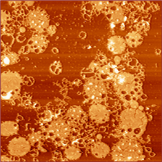
(data) Topographic SFM image of purple membrane patches deposited on mica. Ideally, purple membrane patches are 2D crystals composed primarily of the protein bacteriorhodopsin, are slightly elliptical and of uniform height throughout. The protein here has been modified to change its photoactive properties, yeilding an unexpected significant degradation in stability.
And yet, there is still much that hasn't been done that could be and much that remains to be understood. In the group we work on both of these issues by trying to do things that haven't been done with SPMs and by trying to better understand how they work and what they are telling us.
In the realm of things that haven't been done, we work on SFMs with multiple independently controlled tips. If you have, say, two tips, you can use one to manipulate the sample (via tapping on it, applying a potential, driving current through it, illuminating it via apertureless field enhancement, pulling, pushing, etc.) and one to sense the result, where both the sensing and the manipulation are pointlike. In addition, see the Biophysics section further down the page.
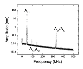
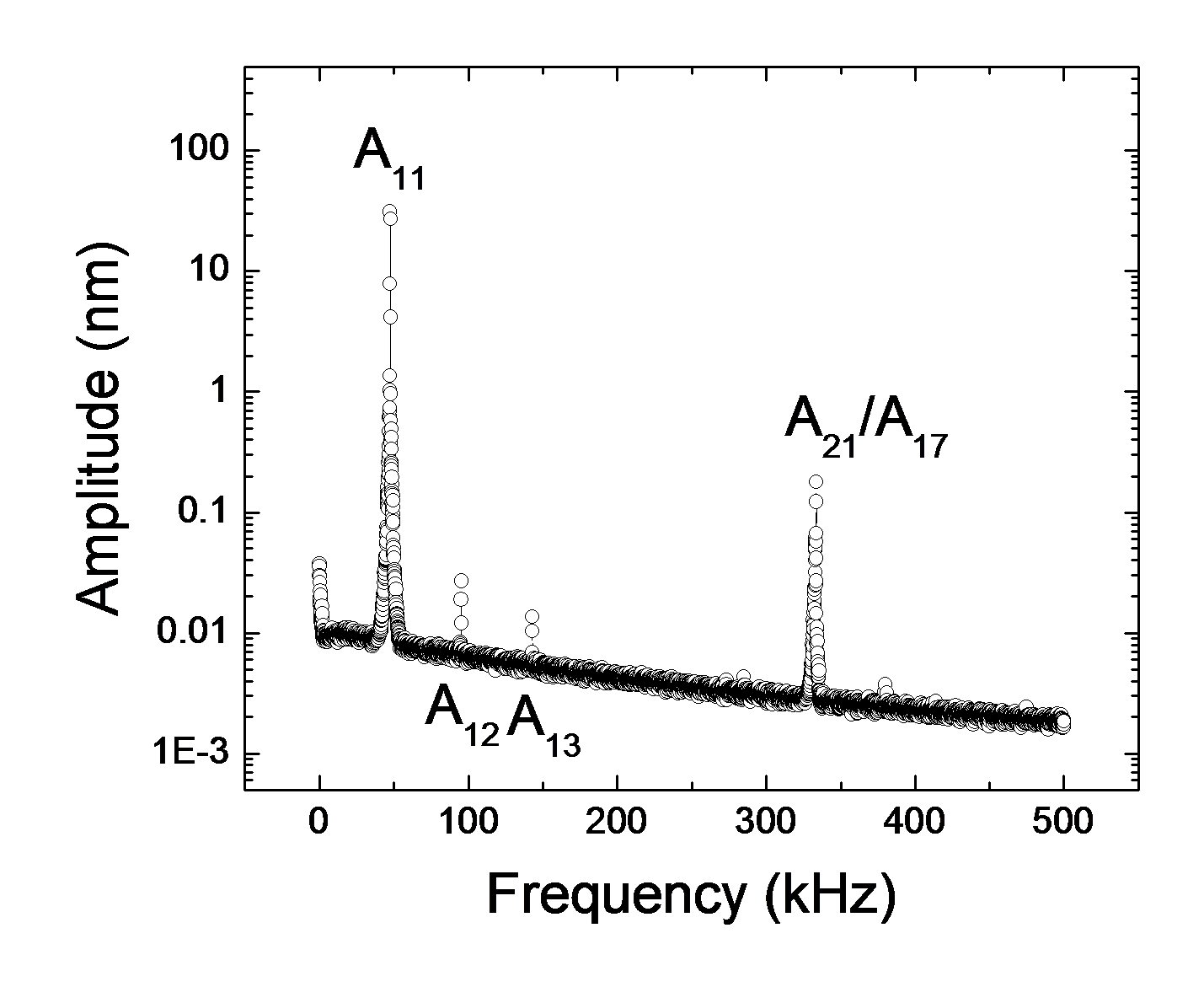
(data) Amplitude spectrum of an SFM cantilever oscillating within imaging distance of a sample. The leftmost peak is at the driving frequency. The second and third harmonics can clearly be seen, as can the second bending mode.
As for increasing the understanding of how SFMs work and what their measurements mean, besides trying understanding the multiprobe system, there is a question of what, exactly, an SFM image is an image of. Many forces act on the cantilever over many interaction distances. The final result is the amplitude, phase, or frequency of the cantilever as a function of position, time, tip-sample separation, etc.. But what is it really?
For instance, the SFM tip-sample interaction force is nonlinear and the cantilever is an extended body, thus in dyanmic mode when the cantilever is driven at some frequency, its motion is nonlinear and contains both harmonics of the driving frequency and higher bending mode oscillations. Many papers have shown these effects and discussed them theoretically yet it's still not clear what they mean. We are attempting to rectify this.
Biophysics
What is the physics of life? What happens far from equilibrium and why? How do complex phenomena emerge from simple ingredients? These are three challenges for condensed matter physics identified by a recent National Academy of Sciences report[3]. Our goal is to approach these questions through the study of individual bacteria. It turns out that many bacteria are capable of producing free electrons as a natural byproduct of their metabolism and many can readily donate those electrons to macroscopic electrodes, thus the current (~1pA/bacterium) should be a relatively rapid (~10s) indicator of metabolic rate.
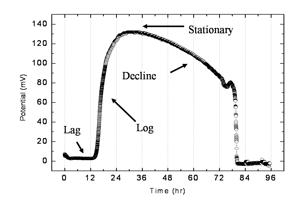
(data) Current (via potential across a resistor) produced by electrogenic bacteria. The shape of the curve follows standard bacterial growth stages.
Combine this with the broad range of nanoscale measurements available with SPM and an array of interesting questions become addressable. To give but one example, some of these organisms appear to accomplish electron donation via long, thin biological structures (pili) which look to be Ohmic conductors over a large potential range[4]. It looks as though Nature may well have already investigated bionanoelectronics; what did it find?
Alternative Energy: Microbial Fuel Cells from Electrogenic Bacteria
Another challenge from the NAS report: How will the energy demands of future generations be met?
Well, how about bacteria that eat just about anything (e.g. Switch Grass, chitin, sewage, pesticides, crude oil, etc.) and turn it into electricity? At 50% efficiency. And room temperature. Without genetically modifying the organisms. Or producing net greenhouse gas emissions.
Join the group, explore the nanoworld, apply physics to bacteria, save the world.
[1] Taken almost verbatim from the Princeton University Biophysics
page.
[2] Chemists and high energy physicists will no doubt rightfully disagree with this statement. Chemists have been working ‘at the bottom’ pretty much since alchemy became chemistry and physicists moved past atoms and electrons round about 1938. U. SC itself has significant Nuclear(1)(2) and High Energy programs.
[3] Most of the six 'challenges' identified in the executive summary of a National Academy of Sciences report, Condensed-Matter and Materials Physics: The Science of the World Around Us.
[4] Extracellular electron transfer via microbial nanowires, Gemma Reguera, Kevin D. McCarthy, Teena Mehta, Julie S. Nicoll, Mark T. Tuominen and Derek R. Lovley, Nature 435, 1098-1101 (23 June 2005) | doi: 10.1038/nature03661.